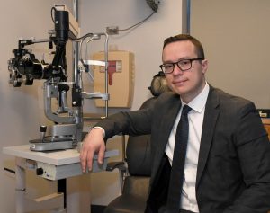
The Education Committee carefully selects presentations from the WGC-2019 for your benefit. This month Gustavo De Moraes introduces the sessions: ANZGS Symposium, Game Changers in Glaucoma and Now you see it, now you don’t: progression or not?.
ANZGS Symposium – The Gillies Lecture, and A Showcase of Glaucoma Research from Australian and New Zealand Universities
In this symposium, leading clinicians and scientists from Australian and New Zealand Universities discuss their most recent work with a focus on blindness prevention, neuro-protection, and risk assessment.
The Gillies Lecture: Of Glaucoma, Prevention of Blindness & the Reverend: A personal Khichdi
Dr. Ravi Thomas describes the prevalence of angle closure and open angle glaucoma in India, as well the rate of conversion from primary angle closure suspect and primary angle closure to angle closure glaucoma. He discussed the impact of cataract extraction on intraocular pressure as well as factors that may predict greater pressure lowering. In addition, he discusses the effect of Vitamin A deficiency and malnutrition on rates of blindness and visual impairment.
Connexin 43 in Retinal Ganglion Cell Injury
Dr. Helen Danesh-Meyer discusses the role of connexins – which are involved in physiology of synapses and gap junctions – in glaucomatous injury. They are also involved in inflammatory responses, which has been shown to play a significant role in glaucoma pathogenesis. In humans, connexin43 protein expression has been shown to increase in the laminar cribrosa and retinal ganglion cell layer of glaucomatous eyes; in addition, glial activation seems to be part of the response to retinal ganglion cells and axonal injury. Finally, she discusses potential neuroprotection therapies based on connexins.
Promoting retinal ganglion cell recovery
Dr. Jonathan Crowston discusses the clinical evidence for visual recovery of retinal ganglion cells in glaucoma. He describes current evidence from glaucoma surgery and the effect of different types of intervention, including physical activity. Finally, he discusses the role of brain-derived neurotrophic factor on retinal ganglion cell recovery.
Novel approaches to neuroprotection – TrkB/Shp2, neuroserpin and RXRs
Dr. Stuart Graham follows up on the previous talk further discussing the current literature on brain-derived neurotrophic factor and neuroprotection of retinal ganglion cells. He then discusses how Shp2-TrkB interactions can affect such relationship, particularly after showing that downregulation of Shp2 is protective against inner retinal functional loss. He ends his talk by addressing the potential role of Shp2 gene therapy in glaucoma by enhancing the BDNF/TrkB pathway.
Bioenergetic Neuroprotetction in Glaucoma
Dr. Robert Casson starts by pointing out the importance of ATP to maintain visual perception, hence suggesting the role of energy delivery for neuroprotection. He then reviews the work on vitamin B3 and Pyruvate and glaucoma neuroprotection in animal models. The closes his talk by discussing the role of nutritional supplements in glaucoma.
Translaminar pressure gradient and glaucoma
Intraocular pressure (IOP) alone does not explain why some patients develop glaucoma or continue to progress despite treatment. Dr. Bill Morgan discusses the importance of translaminar pressure gradient, dependent upon the cerebrospinal fluid pressure, as a key factor to help explain the role of IOP. He shows a series of experiments demonstrating how translaminar pressure gradient affects neural function, blood vessels, and connective tissue in glaucoma.
Machine learning and optic disc assessment for glaucoma
Switching gears to artificial intelligence, Dr. John Grigg presented recent data on how machine learning can help in the detection of glaucoma. In particular, he underscores how machine learning may help ophthalmologists become better at disease detection.
Clinical and molecular risk factors for glaucoma progression and blindness
Dr. James Craig ends the symposium describing the PROGRESSA Study, which aims at stratifying patients based on their risk of progression. He also addresses recent efforts to develop a glaucoma risk score based on genetic markers (the Glaucoma Polygenic Risk Score). He then addresses the role of home IOP monitoring and corneal biomechanics in risk assessment. He concluded by suggesting the need to incorporate multiple markers (genetic and clinical) in generating better tools to predict who are the patients at greatest risk of progressing more rapidly from glaucoma.
Game Changers in Glaucoma
In the opening ceremony of the conference, the speakers discuss recent advances in glaucoma research that are likely to impact the way we diagnose and treat glaucoma in the 21st century.
Machines will make the diagnosis
Dr. Gustavo De Moraes opens the symposium discussing the advances in artificial intelligence and the many ways it may help improve glaucoma management. He underscores the role of the physician ensuring the best use of the output of these machines for clinical management.
So many genes, are we ready for personalized medicine?
Dr. Janey Wiggs then reviews the current knowledge on how genes and genetic variants determine different types of glaucoma. She discusses recent studies based on multisite collaborations that helped detect novel genetic markers of glaucoma. She ends by highlighting the challenges and benefits of collaborations in genetic research.
Who said glaucoma is an incurable disease?
Dr. Keith Martin addresses recent studies suggesting that neuroprotection and neuroregeneration is possible in glaucoma and may translate into novel therapies in the future. He summarizes by suggesting how clinical trials based on pressure-independent therapies are possible and the need for combining different approaches to cure glaucoma.
Detecting stressed ganglion cells
Bridging with the previous talk, Dr. Jonathan Crowston discusses the importance of assessing the stage of retinal ganglion cell dysfunction or death for glaucoma diagnosis as well as developing novel therapies to rescue damaged neurons before it becomes irreversible. With that aim, he describes recent imaging modalities that can detect individual, living retinal ganglion cells; he also discusses previous work suggesting how damage to retinal ganglion cell function assessed with visual fields can be reversible. He suggests it is feasible to image retinal ganglion cells in vivo and improvements in the technique may lead to novel forms of therapy.
Surgical game changers: what’s beyond MIGS?
Dr. Ike Ahmed closes the symposium discussing how being Proactive vs Reactive can influence outcomes in glaucoma surgery. He starts by discussing the rates of progression and blindness in the real world and their possible reasons. He suggests it is now time to change the treatment paradigm in glaucoma by being more aggressive from the start and aiming for lower pressure targets. He ends his talk by proposing minimally invasive procedures and new drug delivery systems as tools to overcome issues with adherence to medical therapy.
Now you see it, now you don’t: progression or not?
Glaucoma is often progressive if left untreated; however, detection of progression remains a central challenge in clinical practice. In this symposium, the speakers address techniques to improve detection of progressive changes and how to differentiate progression from test variability and aging.
Glaucoma Progression or Normal Ageing?
Dr. Fabio Lavinsky opens the symposium by describing the most common methods to detect progression in the clinical setting. He underscores the effect of normal aging as a confounder and reviews the physiologic rate of change based on different clinical tools. He ends his talk by concluding that caution is needed when interpreting age related loss in moderate to advanced glaucoma and stresses how novel imaging tools should help better understand aging effects in-vivo in glaucoma.
Detecting progression in primate glaucoma
Dr. Brad Fortune describes the work of his group on better understanding the sequence of events associated with structural progression in non-human primate glaucoma. He describes the path from optic nerve head deformation and axonal injury all the way down to macular inner retinal thinning and retrograde ganglion cell soma loss, ultimately leading to peripapillary capillary loss.
Advances in VF analysis to detect progression
Switching gears to the importance functional assessment of progression, Dr. Lingam Vijaya discusses how clinical trials in glaucoma assessed visual field progression based on different criteria and compared different analytical methods and parameters that can be employed clinically. She concludes that all portions of the visual field printout should be examined carefully with special attention to test selection and quality.
Role of Macular OCT imaging in clinical glaucoma progression
Dr. Kyung Rim Sung ends the symposium addressing the importance of assessing macular structure and function for enhanced detection of glaucomatous progression. She reviews the importance of macula damage in glaucoma and describes current clinical tools to measure progression. She concludes that macular assessment is complementary to the peripapillary assessment and should be used in combination for better detection of progressive changes in glaucoma.


